Ct Anatomy Pelvis Muscles / Pertinent Ct Anatomy Of The Neck For Interpreting Pet Ct Pet Clinics / The ilium, pubis and ischium.. Almost all of them receive their nerve supply from the posterior (dorsal) rami of spinal nerves and are called the intrinsic group because they act specifically on the vertebral. The sacrum is located inferiorly to the spinal vertebrae, and posteriorly within the pelvis. These bones also act as attachments for many muscles and ligaments within the pelvis and lower limbs. The pelvis's frame is made up of the bones of the pelvis, which connect the axial skeleton to the femurs, and therefore acts in weight bearing of the upper body. These are separated by cartilage at birth and fuse during puberty.
Jul 26, 2021 · the muscles of the thoracic area lie deep to the thoracolumbar fascia, while the muscles of the lumbar area lie between the superficial and middle layers of the fascia. Contraction of the rectus abdominis muscles also compresses abdominal viscera and contributes to forced expiration. These bones also act as attachments for many muscles and ligaments within the pelvis and lower limbs. The floor of the pelvis is made up of the muscles of the pelvis, which support its contents and maintain urinary and faecal continence. Almost all of them receive their nerve supply from the posterior (dorsal) rami of spinal nerves and are called the intrinsic group because they act specifically on the vertebral.
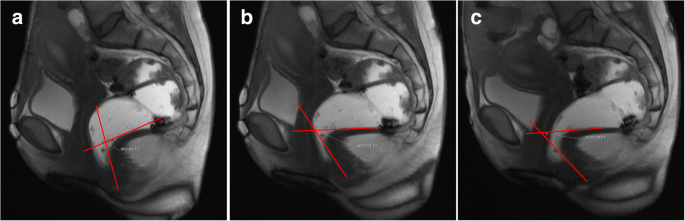
Dynamic Magnetic Resonance Imaging Of The Female Pelvic Floor A Pictorial Review Insights Into Imaging Full Text from media.springernature.com Contraction of the rectus abdominis muscles also compresses abdominal viscera and contributes to forced expiration. The talus is the bone that makes up the lower part of the ankle joint. The sacrum is located inferiorly to the spinal vertebrae, and posteriorly within the pelvis. These are separated by cartilage at birth and fuse during puberty. We would like to show you a description here but the site won't allow us. These nerves control your muscles so that you can walk, blink, swallow, pick things up and do other activities. Contrast material may be injected into a vein or the spinal fluid to enhance the scan. The floor of the pelvis is made up of the muscles of the pelvis, which support its contents and maintain urinary and faecal continence.
These bones also act as attachments for many muscles and ligaments within the pelvis and lower limbs.
The floor of the pelvis is made up of the muscles of the pelvis, which support its contents and maintain urinary and faecal continence. These are separated by cartilage at birth and fuse during puberty. The sacrum is located inferiorly to the spinal vertebrae, and posteriorly within the pelvis. Peripheral nerve tumors can occur anywhere in the body. Ct images are available in 3 different planes (transverse, sagittal and dorsal) with two kinds of contrast (bones/lungs and soft tissues/mediastinum/vessels). Almost all of them receive their nerve supply from the posterior (dorsal) rami of spinal nerves and are called the intrinsic group because they act specifically on the vertebral. Contraction of the rectus abdominis muscles also compresses abdominal viscera and contributes to forced expiration. Contrast material may be injected into a vein or the spinal fluid to enhance the scan. Jul 26, 2021 · the muscles of the thoracic area lie deep to the thoracolumbar fascia, while the muscles of the lumbar area lie between the superficial and middle layers of the fascia. Jun 17, 2021 · the muscles are also involved in stabilising the pelvis and controlling the neutral position of the pelvis, and preventing abnormalities in the pelvic position, such as anterior or posterior pelvic tilt. The pelvis's frame is made up of the bones of the pelvis, which connect the axial skeleton to the femurs, and therefore acts in weight bearing of the upper body. We would like to show you a description here but the site won't allow us. The ilium, pubis and ischium.
The sacrum is located inferiorly to the spinal vertebrae, and posteriorly within the pelvis. These nerves control your muscles so that you can walk, blink, swallow, pick things up and do other activities. We would like to show you a description here but the site won't allow us. The ilium, pubis and ischium. These bones also act as attachments for many muscles and ligaments within the pelvis and lower limbs.
Module 5 Pelvis Imaging from hca.fujifilm.com These are separated by cartilage at birth and fuse during puberty. We would like to show you a description here but the site won't allow us. Jul 15, 2020 · peripheral nerve tumors are growths in or near the strands of tissue (nerves) that transmit signals from your brain to the rest of your body. These nerves control your muscles so that you can walk, blink, swallow, pick things up and do other activities. The hip bone has three parts: Contrast material may be injected into a vein or the spinal fluid to enhance the scan. Contraction of the rectus abdominis muscles also compresses abdominal viscera and contributes to forced expiration. The floor of the pelvis is made up of the muscles of the pelvis, which support its contents and maintain urinary and faecal continence.
The hip bone has three parts:
The sacrum is located inferiorly to the spinal vertebrae, and posteriorly within the pelvis. Jul 26, 2021 · the muscles of the thoracic area lie deep to the thoracolumbar fascia, while the muscles of the lumbar area lie between the superficial and middle layers of the fascia. The pelvis's frame is made up of the bones of the pelvis, which connect the axial skeleton to the femurs, and therefore acts in weight bearing of the upper body. These nerves control your muscles so that you can walk, blink, swallow, pick things up and do other activities. The floor of the pelvis is made up of the muscles of the pelvis, which support its contents and maintain urinary and faecal continence. Contraction of the rectus abdominis muscles also compresses abdominal viscera and contributes to forced expiration. Jul 15, 2020 · peripheral nerve tumors are growths in or near the strands of tissue (nerves) that transmit signals from your brain to the rest of your body. The talus is the bone that makes up the lower part of the ankle joint. Almost all of them receive their nerve supply from the posterior (dorsal) rami of spinal nerves and are called the intrinsic group because they act specifically on the vertebral. Ct images are available in 3 different planes (transverse, sagittal and dorsal) with two kinds of contrast (bones/lungs and soft tissues/mediastinum/vessels). The hip bone has three parts: These are separated by cartilage at birth and fuse during puberty. Peripheral nerve tumors can occur anywhere in the body.
Jul 15, 2020 · peripheral nerve tumors are growths in or near the strands of tissue (nerves) that transmit signals from your brain to the rest of your body. Peripheral nerve tumors can occur anywhere in the body. The hip bone has three parts: Contraction of the rectus abdominis muscles also compresses abdominal viscera and contributes to forced expiration. The ilium, pubis and ischium.
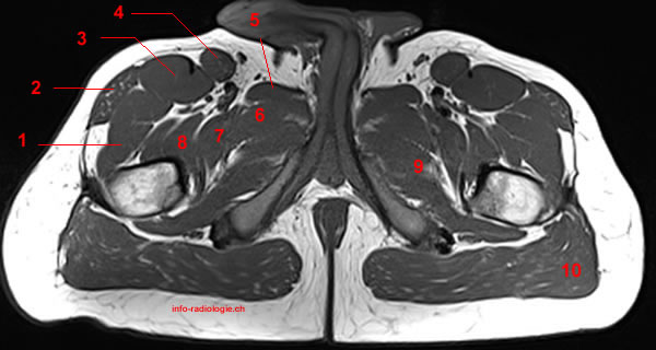
Mri Of The Thigh Detailed Anatomy Superior Part W Radiology from w-radiology.com The ilium, pubis and ischium. Peripheral nerve tumors can occur anywhere in the body. Jul 15, 2020 · peripheral nerve tumors are growths in or near the strands of tissue (nerves) that transmit signals from your brain to the rest of your body. The pelvis's frame is made up of the bones of the pelvis, which connect the axial skeleton to the femurs, and therefore acts in weight bearing of the upper body. These nerves control your muscles so that you can walk, blink, swallow, pick things up and do other activities. Ct images are available in 3 different planes (transverse, sagittal and dorsal) with two kinds of contrast (bones/lungs and soft tissues/mediastinum/vessels). Jun 17, 2021 · the muscles are also involved in stabilising the pelvis and controlling the neutral position of the pelvis, and preventing abnormalities in the pelvic position, such as anterior or posterior pelvic tilt. Because the talus is so important for ankle movement, a fracture often results in substantial loss of motion and function.
Almost all of them receive their nerve supply from the posterior (dorsal) rami of spinal nerves and are called the intrinsic group because they act specifically on the vertebral.
Jul 26, 2021 · the muscles of the thoracic area lie deep to the thoracolumbar fascia, while the muscles of the lumbar area lie between the superficial and middle layers of the fascia. Almost all of them receive their nerve supply from the posterior (dorsal) rami of spinal nerves and are called the intrinsic group because they act specifically on the vertebral. These bones also act as attachments for many muscles and ligaments within the pelvis and lower limbs. We would like to show you a description here but the site won't allow us. Contraction of the rectus abdominis muscles also compresses abdominal viscera and contributes to forced expiration. The hip bone has three parts: Because the talus is so important for ankle movement, a fracture often results in substantial loss of motion and function. The talus is the bone that makes up the lower part of the ankle joint. The ilium, pubis and ischium. The floor of the pelvis is made up of the muscles of the pelvis, which support its contents and maintain urinary and faecal continence. The pelvis's frame is made up of the bones of the pelvis, which connect the axial skeleton to the femurs, and therefore acts in weight bearing of the upper body. These are separated by cartilage at birth and fuse during puberty. Contrast material may be injected into a vein or the spinal fluid to enhance the scan.
Contraction of the rectus abdominis muscles also compresses abdominal viscera and contributes to forced expiration anatomy muscles pelvis. Jul 15, 2020 · peripheral nerve tumors are growths in or near the strands of tissue (nerves) that transmit signals from your brain to the rest of your body.


0 Komentar